Echinococosis
Echinococcosis is a zoonotic disease caused by larval stages of the dog tapeworm. The definitive natural host of this tapeworm is dogs but the disease occurs when humans become the accidental host of the Echinococcus granuloses larval stage.
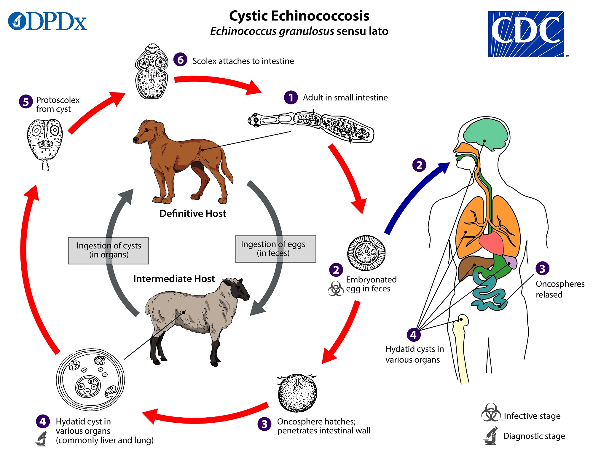
In the natural lifecycle of this species the larval and adult stages revolves around the dog and the sheep as shown in this great image I found in CDC website.
There are a few types of echinococcosis
Cystic Echinococcosis : Hydatid cysts (Echinococcus granulosis)
Alveolar Echinococcosis: Lungs (Echinococcus multiocularis)
Neotropical Echinococcosis: (Echinococcus vogeli)
Uni-cystic Echinococcosis: (Echinococcus oligarthrus)
Morphology
This worm has 3 segments (or 4 segments including the scolex)
Scolex
Immature Segment
Mature Segment
Gravid Segment
Lifecycle Stages in Humans
Embryonated eggs in the faeces of dogs.
Oncosphere hatches and penetrates the intestinal wall.
Hydatid cyst in the liver lungs etc.
Here the cystic lifecycle ends. A canine host is needed to continue the lifecycle beyond the hydatid cyst stage.
Hydatid Cyst
Hydatid cyst is a complex structure it is surrounded by a host collagen capsule. A laminated layer and a germinal layer. Germinal layer generates daughter cells, and they contain Proto scolex.
Daughter cyst rupture and generate sand like structure collected at the bottom of the cyst. This is called hydatid sand, and it can disseminate and infect other parts of the body.
CT, MRI and ultrasound scans and serological diagnosis can be used to identify the parasite. ELISA will need a confirmation with a western blot.
Casoni’s skin test can be used to diagnose this disease.
Treatment
Management of the hydatid cyst is needed. Before surgery Albendazole is used as the drug of choice to prevent dissemination. Removal of cyst fluid and fill the space with Silver Nitrate and 10% formalin for 10 minutes is also done. This will kill the germinal membrane.
After that it becomes safe for a surgeon to cut and drain the cyst. The germinal membrane also needs to be removed while doing so.
Comments
Join the discussionNo comments yet. Be the first to comment.
